In this section, Prof G Sivakumar lists down the clinical signs of Diabetic Foot syndrome.
Diabetic Foot Syndrome
Identifying long-term complications of diabetes and preventing ulceration in the foot.
Neuropathy is common in 30 to 40 % of patients with diabetic mellitus. 20% of them develop foot ulcers.
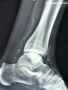
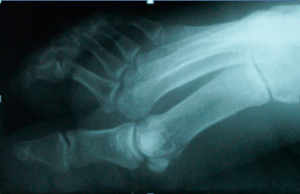
These are clinical and radiological signs that foretell the ulceration.
These are referred as GRADE – O lesions.
- “Intrinsic minus” deformity due to wasting of small muscles.

- Neuropathic joint is common and is called “CHARCOT”joint.
- “Monckeberg’s sclerosis” of the Tibial arteries is due to calcification of the Tunica media.

- Prominent leg veins in recumbent posture, called JD Ward sign. This is due to increased venous flow due to a-v shunting of blood at the capillary level due to neuropathy.
- Hyper extension at First metatarso – phalyngeal joint. This deformity is common in the development of plantar ulcers.

- Collapse of foot arches.
- Fractures and dislocations.
- Osteopaenia.
- Unaware of foreign bodies.
- Pedal edema.
- Absent pedal arterial pulses.
- Plantar sites of peaking pressure points & callosities.
- Bunions & bursae.
- Reduced Vibration sensation.
- Gas shadows in skiagram, signs of impending ulceration.
- Hallux valgus & rigidus.
“Ulceration in the foot in diabetics is predictable and preventable”
